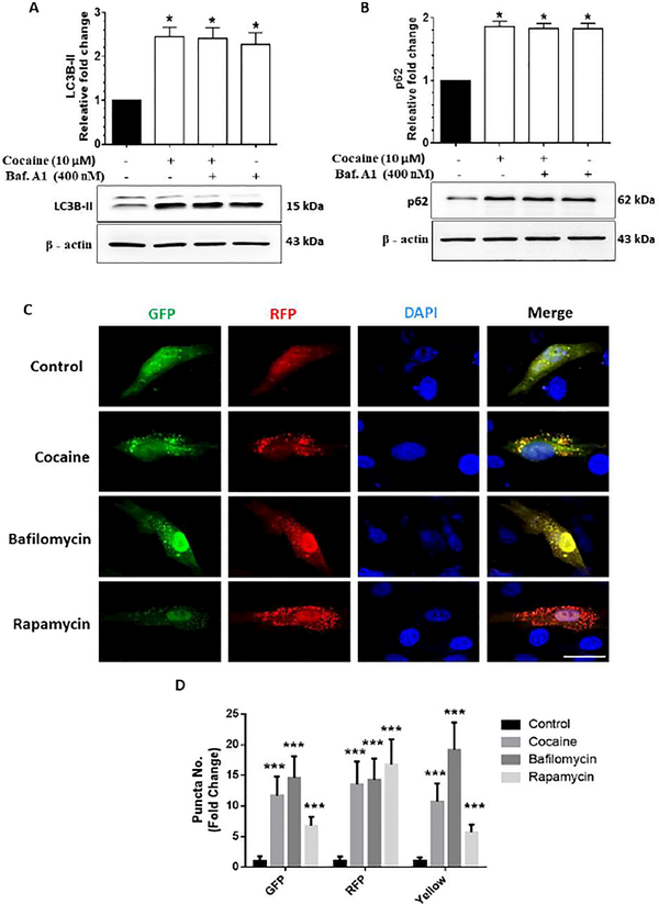
Figure 3.
Cocaine-mediated dysregulated autophagy in human primary pericytes (HBVP). Representative western blots showing protein levels of LC3B-II (A) and p62 (B) in human HBVP exposed to cocaine (10 μM) for 12 h followed by treatment with 400 nM BAF, which was added in the last 4 h of the 12 h treatment period. (C) Representative fluorescent photomicrographs showing the LC3B-II puncta formation in HBVP cells transfected with tandem fluorescent-tagged LC3B-II plasmid and treated with 10 μM cocaine for 12 h, 400 nM BAF - last 4 h of the 12 h treatment period and 10 nM rapamycin for 24 h. Quantitative analyses of yellow, red and green puncta (D) formation in different experimental groups of tandem fluorescent-tagged LC3B-II plasmid transfected HBVP. Scale bar: 10 μm. β-actin was used as a loading control for all experiments. Data are presented as mean ± SEM; the mean is derived from 6 independent experiments (n = 6). Abbreviation: Baf. A1: Bafilomycin A1. One-way ANOVA followed by Bonferroni post hoc test was used to determine the statistical significance. *, P<0.05 vs control.

