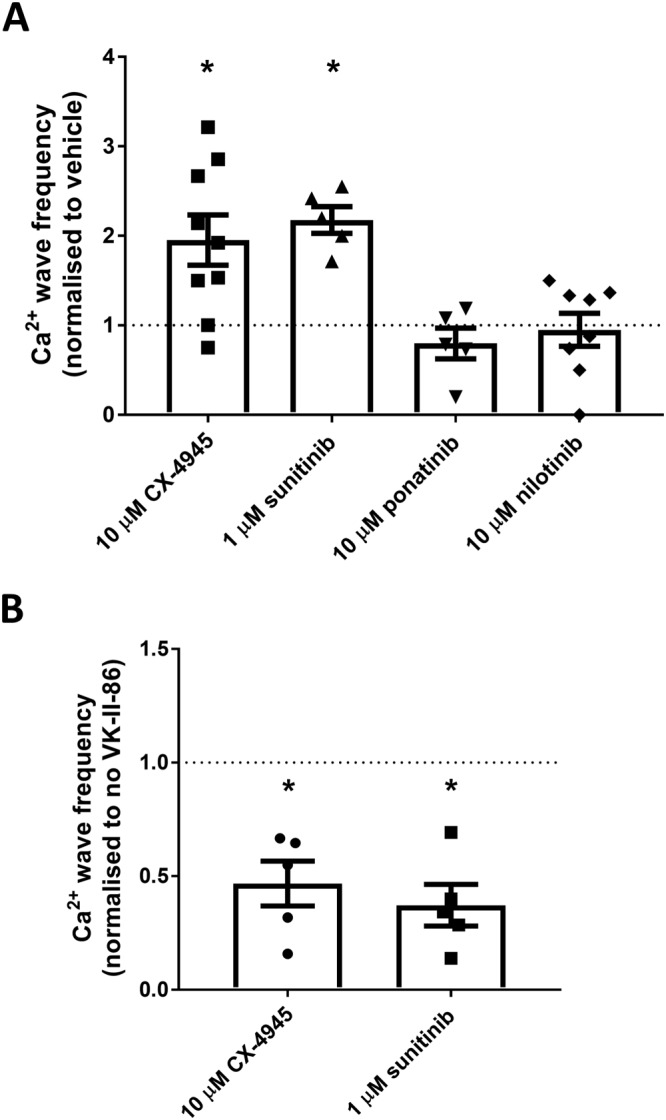
Figure 7.

Class I kinase inhibitors increase Ca2+ wave generation in rat cardiomyocytes which can be prevented by VK‐II‐86. Freshly isolated rat ventricular myocytes were loaded with fluo‐4 in KRH buffer. (A) Cells were superfused with KRH containing 1 mM Ca2+ and paced at 0.5 Hz. Cells were treated with vehicle or 10 μM of CX‐4945, ponatinib or nilotinib or 1 μM sunitinib for 10 min before pacing was stopped for 2 min. The number of spontaneous Ca2+ waves occurring during the 2 min was recorded. At the end of each experiment, cells were perfused with 20 mM caffeine to confirm cell viability. Data shown are the Ca2+ wave frequency (mean ± SEM) normalized to vehicle‐treated cells from the same animal (10 μM CX‐4945 n = 7; 1 μM sunitinib n = 5; 10 μM ponatinib n = 7; 10 μM nilotinib n = 8, different animals); *P < 0.05 versus vehicle. (B) As with panel (A) except cells were pre‐incubated with 1 μM VK‐II‐86 or vehicle control (DMSO) for 30 min prior to imaging. Data shown are the Ca2+ wave frequency (mean ± SEM) normalized to non‐VK‐II‐86‐treated cells (vehicle control) from the same animal (n = 5 different animals); *P < 0.05 versus vehicle.
