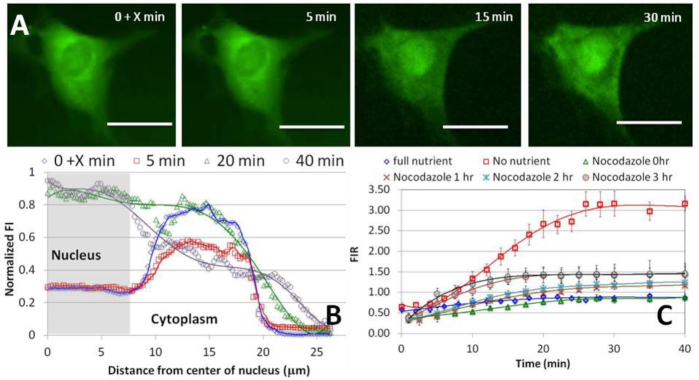
Figure 8.
(A) rhd-tRNA emission intensity from a 0.4 μm confocal slice of a cell pre-exposed for 3 h to 100 nM extracellular microtubule de-polymerizing agent nocodazole. Scale bar 25 μm. X < 30 s corresponds to time after injection. See SI-S7 for additional data. (B) Fluorescent intensity along the cell's diameter at various times after rhd-tRNA injection into the cell. (C) Fluorescent intensity ratio (FIR) as a function of time under 100% and 0% nutrition in the absence of nocodazole, and under 100% nutrition and 0.02 (N = 11), 1 (N = 7), 2 (N = 8), 3 (N = 6) and 4 (N = 6) hours pre-exposure to nocodazole.

