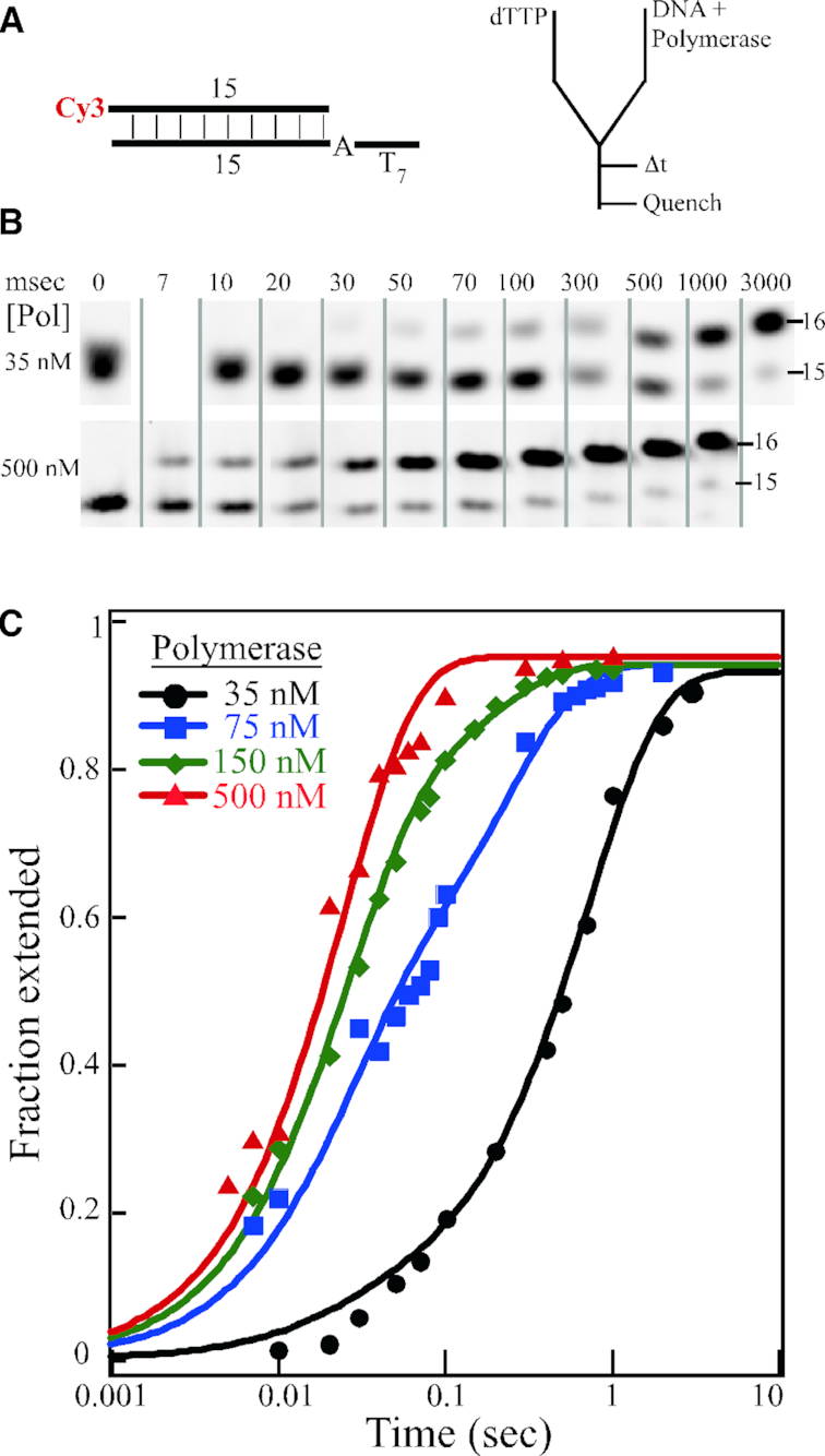
Figure 2.

Single nucleotide extension studies. (A) DNA substrate and rapid quench experimental setup. (B) Selected time points of single nucleotide extension by 35 and 500 nM mini-Pol δ-DV, pre-incubated with 50 nM 23/15 template/primer DNA. Reactions were initiated with 250 μM final dTTP. The full gels are shown in Supplementary Figures S4A and S4B. (C) Quantification of the data for 35 nM (black), 75 nM (blue), 150 nM (green) and 500 nM (red) mini-Pol δ-DV. The fractional extension of the 15-mer product is plotted against time on a logarithmic scale.
