Figure 4.

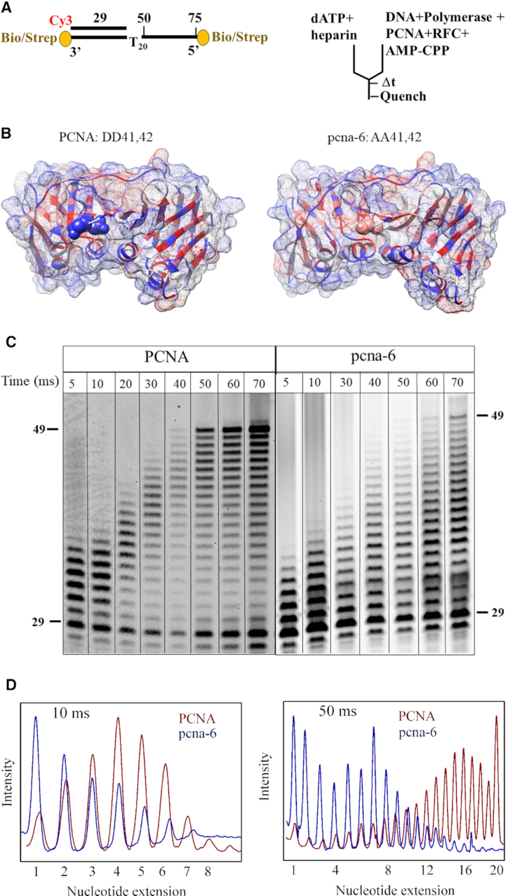
Replication defects of pcna-6 (DD41,42AA). (A) DNA substrate and rapid quench experimental setup (as in Figure 3, except for the addition of a heparin trap). (B) Crystal structure of wild-type PCNA (1PLR) with D41 and D42 shown as CPK in blue and pcna-6 (5V7K) with A41 and A42 shown in pink. Hydrophilic surface in blue and hydrophobic surface in red. (C) Time courses of primer extension by mini-Pol δ-DV (300 nM) pre-incubated with 50 nM DNA in the presence of 75 nM RFC and 100 μM AMP-CPP, and either 150 nM PCNA (left), or 150 nM pcna-6 (right). Reactions were initiated with 250 μM final dATP and 17 μg/ml final heparin sulphate. Complete data are in Supplementary Figures S4E and S4F. (D) Comparison of product distribution with PCNA (red) and pcna-6 (blue) at 10 and 50 milliseconds. Normalized band intensities are shown with the highest intensity for each distribution set to 1.
