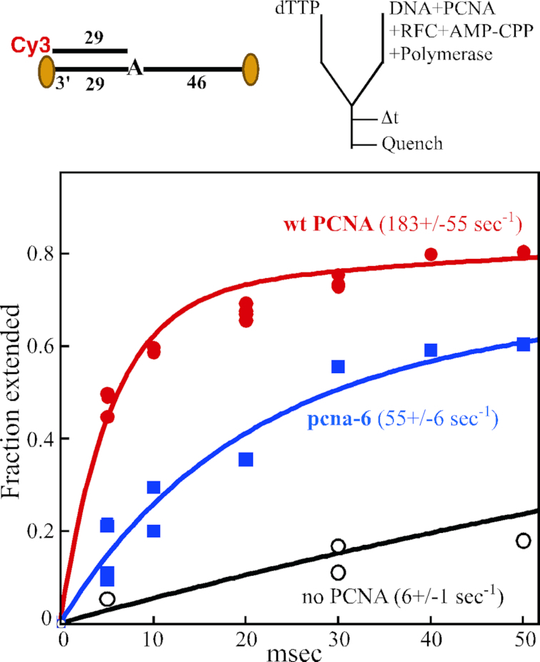
Figure 5.

Single-nucleotide extension defect of pcna-6 (DD41,42AA) at 12°C. Top, substrate and experimental setup are identical to described in Figure 3, except that the 76/29 substrate has a single template adenine residue, and we used full-length Pol δ-DV. Upon addition of dTTP to the preformed complex, extension is by one single nucleotide. Assays were performed at 12°C. Bottom, primer extension is plotted against time in the absence of PCNA (black), with wild-type PCNA (red) or with pcna-6 (blue). The data were modeled to the sum of two exponentials, and kfast is shown (see Materials and Methods). For the assay without PCNA, the assay was taken out to 1 s, at which time 84% extension had occurred. These data were included in the modelling, but only the portion up to 50 ms is shown.
