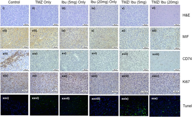
Figure 6.
Immunohistochemistry and immunofluorescence analysis of excised mouse brain derived from the in vivo experiment. Brains from the treated mice (n = 8 per treatment group) were harvested at the time of euthanasia. Sections (4 μm) were cut and mounted on ultrafrost slides. H&E, MIF, CD74 and Ki67 proteins were assessed with IHC while Tunel positivity was assessed with immunofluorescence (IF). Representative photomicrographs are presented from each treatment group (i–iv H&E; vii–xii MIF; xiii–xviii CD74; xix–xxiv Ki67 and xxv–xxx Tunel). All images were taken at a magnification of x20 and the error bars represent 100 μm.

