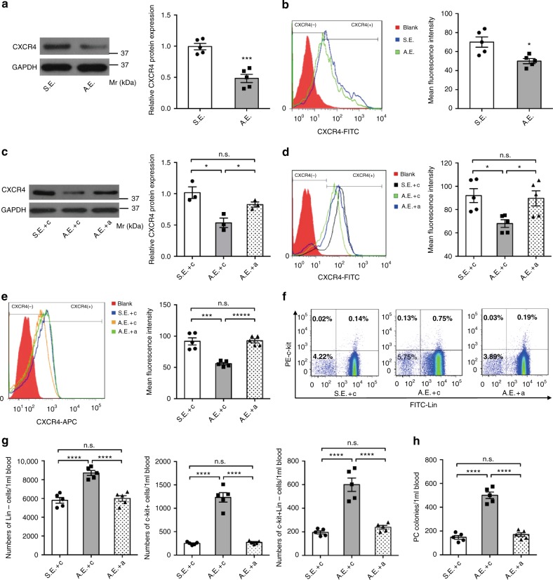
Fig. 4.
Exosomal transfer of myo-miRs downregulates CXCR4 in BM-MNCs and induces BM PC mobilization. Exosomes were isolated from the plasma of AMI mice (AMI-exosomes, A.E.) or Sham mice (Sham-exosomes, S.E.) 6 h post-surgery. a–d In vitro, BM-MNCs (2 × 10⁷/well) were cultured for 24 h with A.E. (20ug), S.E. (20 μg), A.E. plus 10 nM non-targeting scrambled control sequence (c) (A.E. + c), S.E. + c, or A.E. plus 10 nM (4 × 2.5 nM each) antagomir (A.E. + a), then quantified for CXCR4 expression by Western-blotting (a, c; n = 5 biologically independent samples per group for a, n = 3 biologically independent samples per group for c) and flow-cytometry (b, d, n = 5 biologically independent samples per group). *p < 0.05, ***p < 0.001. e–h In vivo, C57BL/6 mice were injected with a (80 mg [20 mg each] antagomir/kg body weight/day) or c (80 mg/kg/day) for 3 consecutive days, then subjected to AMI or Sham surgery and 6 h later, to isolation of plasma exosomes. The isolated exosomes were subsequently i.v. injected into intact C57BL/6 mice (40 μg diluted in 300 μL PBS/mouse) and 12 h later, BM-MNCS and PB-MNCs were isolated from the recipient mice for assessments of PC mobilization. e–g Flow cytometry analyzes for CXCR4 expression in BM-MNCs (e), percentages of c-kit+, Lin–, and c-kit+Lin– cells in the PB-MNCs (f), and calculation of absolute c-kit+, Lin–, and c-kit+Lin– cell numbers per 1 ml PB (g). h The colony-forming PCs in the PB-MNCs were evaluated via colony formation assay. ***p < 0.001, ****p < 0.0001, n.s., no significant. n = 5 animals per group. An unpaired t test was used in (a, b) and a one-way ANOVA was used in (c–h) for statistical analysis. Error bars represent mean ± s.e.m

