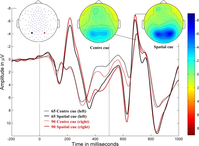
Figure 2.
Orienting. Grand-averaged ERP waveforms for the spatially-cued stimulus (solid lines) and centre-cued stimulus (dotted lines) at posterior electrodes (90, red, right hemisphere; 65, black, left hemisphere) in typically developing children (negativity up). Cue onset at 0 ms and target stimulus onset at 500 ms. Amplitude topographies for spatially-cued and centre-cued target stimuli at 686 ms (i.e., 186 ms after target stimulus onset).

