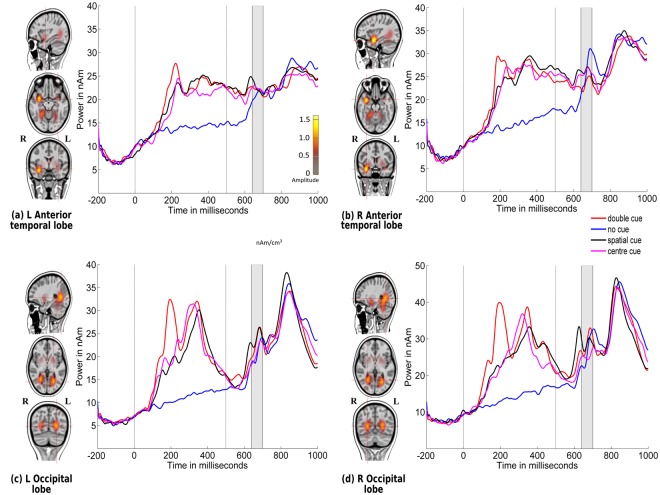
Figure 5.
Source locations of grand-averaged ERPs collapsed across all conditions (congruent and incongruent stimuli: no cue, double cue, centre cue, and spatial cue) over time points of the N1 period of the target stimulus (140–200 ms) using CLARA in typically developing children. Cue onset is at 0 ms and target stimulus onset is at 500 ms. Brain activations were localized in the (a) left anterior temporal lobe, (b) right anterior temporal lobe, (c) left occipital lobe, and (d) right occipital lobe. Grand-averaged source waveforms for double-cued (red), non-cued (blue), spatially-cued (black), and centre-cued (magenta) stimuli, extracted using regional sources at the foci revealed by CLARA, are shown on the right side of each source. The colour bar denotes source amplitude. The shaded grey area denotes the source analysis time window.

