Figure 1.
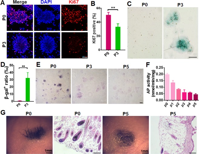
Senescence and hair induction capacity of SKPs in culture. (A,B) Passage 0 and 3 SKPs were immunostained for Ki67. Nuclei were stained with DAPI. Scale bar, 50 μm (A). The percentages of Ki67-positive SKPs were quantified by Image J (B). Data are represented as means ± SEM (n = 3; ***P < 0.001). At least 200 cells were counted in each experiment. (C,D) SKPs in different passages (P) were stained for SA-β-galactosidase (Green) for cellular senescence (C, Scale bars: 100 μm) and the percentage of cells positive for SA-β-galactosidase were counted using Image J (D, mean ± SEM; n = 3; ***P < 0.001) (D). (E,F) Representative images of SKP spheres in P0, 3 and 5 after AP stain (E, Scale bars: 50 μm). AP activity of SKPs in P0-5 were analyzed using an AP assay kit (F) (mean ± SEM; n = 3; ***P < 0.001). (G) Hair genesis of SKPs in different passages. SKPs in P0 and P5 were implanted into excisional wounds of nude mice in combination with freshly isolated neonatal mouse epidermal cells. Twenty days post transplantation, the wound site was photographed under a dissecting microscope and hairs generated were shown. Scale bars: 50 μm.

