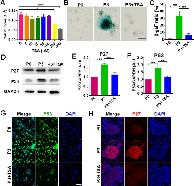
Figure 2.
TSA alleviates SKP senescence. (A) SKPs in culture were treated with TSA at concentrations of 0, 5, 25, 50, 100, 200 and 400 nM for 24 h and cell numbers were counted (mean ± SEM; n = 3; *P < 0.05; ***P < 0.001). (B,C) SKPs in passage (P)0 or P3 treated without or with 100 nM TSA were stained for SA-β-galactosidase (green) and a representative image for each group was shown (B, Scale bars: 100 μm). The percentages of β-gal-positive SKPs were quantified using Image J (C). Data are represented as a mean ± SEM (n = 3; **P < 0.01). (D–F) The cells were analyzed by Western blotting with antibodies recognizing P53, P27 and GAPDH (as a loading control), respectively (D), and the intensity of bands of P27 (E) and P53 (F) was quantified by densitometry and normalized to GAPDH (mean ± SEM; n = 3; **P < 0.01; ***P < 0.001). (G,H) SKPs in P0 and P3 treated with or without 100 nM TSA were analyzed by immunofluorescence staining with anti-P53 (G) or anti-P27 (H) antibody. Nuclei were stained with DAPI. Scale bars: 50 μm.

