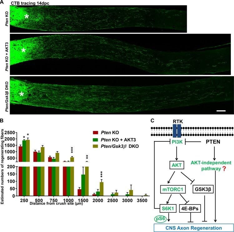
Fig. 4. AKT3 overexpression or GSK3β deletion further increases PTEN KO-induced axon regeneration.
a Confocal images of ON longitudinal sections showing regenerating fibers labeled with CTB-Alexa 488 2 weeks after ON crush. Scale bar, 100 µm. *Crush site. b Quantification of regenerating fibers at different distances distal to the lesion site. Data are presented as means ± s.e.m., n = 11–13. *p < 0.05, **p < 0.01, ***p < 0.001 versus Pten KO alone. One-way ANOVA with Bonferroni’s post hoc test. c A schematic illustration of AKT-dependent and -independent pathways downstream of PTEN deletion that act additively to promote CNS axon regeneration. PTEN deletion minimally activates AKT due to the feedback inhibition of mTORC1/S6K1. The low AKT activation results in adequate mTORC1 activation and low GSK3β inhibition, which are necessary for axon regeneration. The question mark represents unknown effectors of PTEN deletion, which are AKT independent and cooperate with AKT-dependent signaling to promote axon regeneration. ANOVA, analysis of variance; CTB, cholera toxin β; ON, optic nerve

