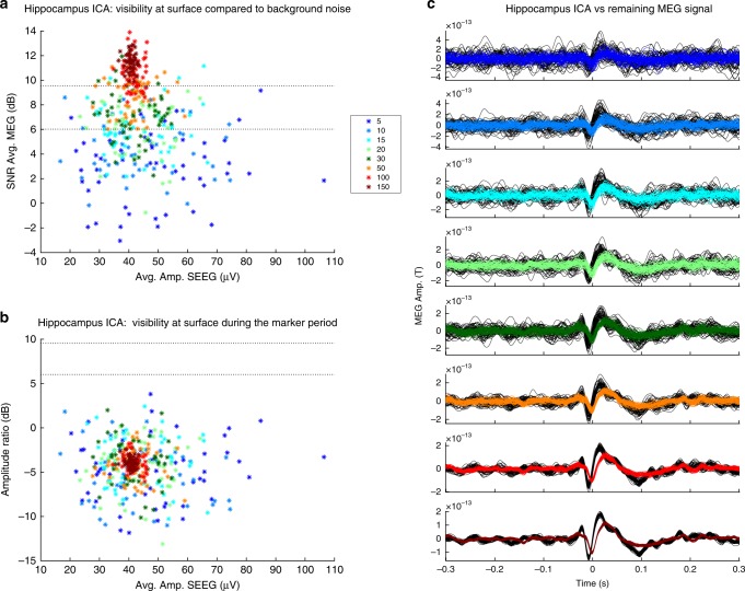
Fig. 4.
SNR evaluation of a ICA component correlated with hippocampus (“Hippocampus ICA”) in the “continuous analysis”. a ICA visibility compared to background noise: the stars indicate the number of markers to average to obtain a certain visibility (SNR). b ICA visibility during the marked period. c Average time course of the ICA component in relationship to the MEG signal on the sensor where the ICA topography has the maximal absolute value. Each color line refers to the number of events that had been averaged (refers to figure legend)

