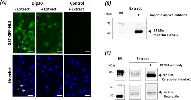
Figure 5.
Nuclear import of the fusion protein GST-GFP-NLS in the permeabilized cells. Cells permeabilized with 30 µg/mL digitonin (Dig30) and non-permeabilized cells (Control) were incubated with GST-GFP-NLS in the absence (−Extract) or presence (+Extract) of Xenopus egg extract supplemented with ATP for 1 h at 25 °C. The incorporation of GST-GFP-NLS was assessed by green fluorescence. Cell nuclei were counterstained with Hoechst 33243. Note that without egg extract, the nuclei are not labelled (arrows). Due to light scattering of the fluorescence in the cytoplasm, the nuclei appear smaller than they are. The egg extract restored nuclear import in the permeabilized cells. These pictures are representative of five independent replicates. Scale bar = 20 µm. Detection of importin alpha-1 (55 kDa) (B) and Karyopherin beta-1 (KPNB1–97 kDa) (C) in Xenopus egg extract by western blot in the presence (+) or in absence (−) of the anti-Xenopus importin alpha-1 antibody (clone 15) and the anti-rat KPNB1 antibody (KPNB1-clone 23). Beta actin was used as a loading control. M: size markers. The cropped blots came from the same gels and were analyzed with the same exposure times (B:1 sec; C:1 min).

