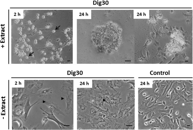
Figure 7.
Morphology of the permeabilized cells over the time post-resealing. Permeabilized cells (Dig30) were treated with Xenopus egg extract (+Extract) or left without (−Extract) for 1 h at 25 °C. Cells were then incubated in the resealing medium for 2 h. Permeabilized cell behavior was assessed by phase contrast microscopy after the 2 h resealing and 24 h post-resealing. The permeabilized cells that were egg-extract treated exhibited a round and refracting morphology (arrows) that progressively spread. By contrast, the permeabilized cells that were not exposed to the Xenopus egg extract did not survive in the resealing medium (arrow heads). These pictures are representative of six independent replicates. Non-permeabilized cells incubated in culture medium were included as control cells. Scale bar = 20 µm.

