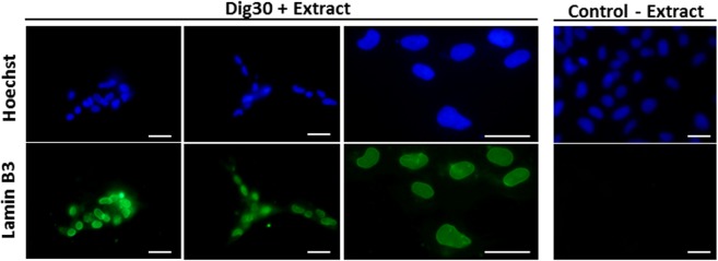
Figure 8.
Detection of Xenopus Lamin B3 in treated cells 24 h post-resealing. Permeabilized cells were treated with Xenopus egg extract (Dig30 + Extract) for 1 h at 25 °C and incubated in resealing medium for 2 h. At 24 h post-resealing, Lamin B3 was detected by immunofluorescence in the nuclei of recovered cells. Nuclei were counterstained with Hoechst 33243. Several representative fields are shown including an enlarged view. Non-permeabilized cells were incubated in absence of Xenopus egg extract (Control – Extract). These pictures are representative of three independent replicates. Scale bar = 20 µm.

