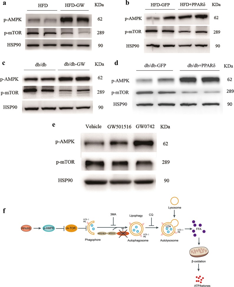
Fig. 8. The induction of PPARδ-induced FAO mediated by autophagy-lysosomes through AMPK/mTOR pathway.
The phosphorylation levels of AMPK and mTOR in vivo and in vitro. a HFD and HFD + GW501516 mice (n = 4–6 per group). b HFD + GFP and HFD + PPARδ mice (n = 4–6 per group). c db/db and db/db + GW501516 mice (n = 4–6 per group). d db/db + GFP and db/db + PPARδ mice (n = 4–6 per group). e PMH were treated with the PPARδ ligand GW501516 (1 μM) or GW0742 (1 μM) for 24 h. Three independent experiments were performed. f PPARδ can increase the phosphorylation of AMPK, and the nutrient-sensing kinase mTOR is inhibited by AMPK. Autophagy is known to be promoted by AMP, so the inhibition of mTOR may activate autophagy through the PPARδ/AMPK/mTOR pathway. Autophagosomes fuse with lysosomes to form mature autolysosomes, in which engulfed contents, including cytosolic lipid droplets (LDs), are degraded. After autolysosome formation, the breakdown of TGs in LDs generates FFAs, which can undergo β-oxidation for ATP and ketone production. PPARδ can activate autophagy, thus increasing LD degradation and hepatic FAO. Two widely used autophagy inhibitors, 3-MA and CQ, and ATG5 KD can also block the effects of PPARδ. The diagram shows that PPARδ has a beneficial effect in the reduction in intrahepatic lipid content and stimulates β-oxidation by an autophagy–lysosomal pathway

