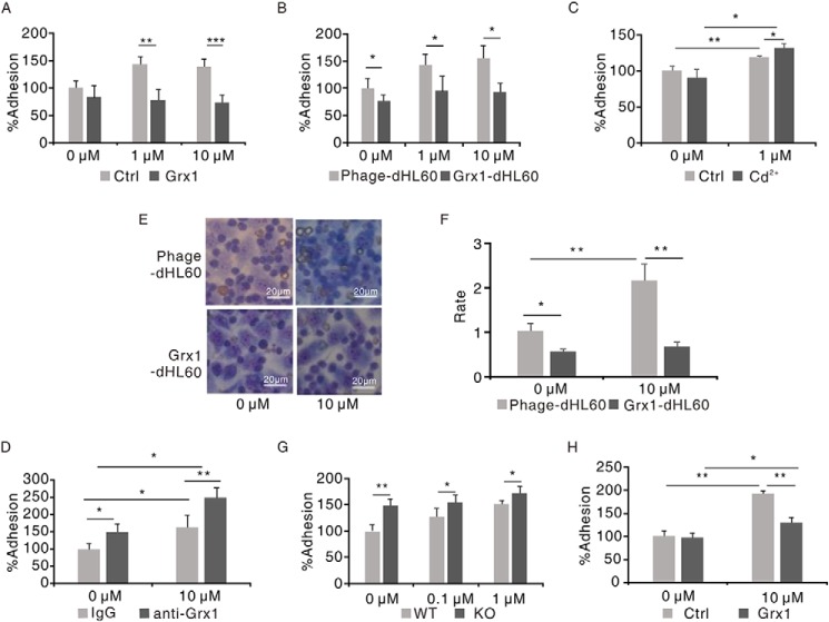
Figure 5.
Grx1 suppressed the adhesion of neutrophils to endothelial cells. A, CFSE-labeled dHL60 cells were preincubated with Grx1 protein (10 μg/ml) or not for 30 min and then stimulated with the indicated concentrations of fMLF and allowed to adhere to HUVECs. The adhesion of cells without Grx1 incubation and stimulation was defined as 100%. **, p < 0.01; ***, p < 0.001 versus control (Ctrl). B, CFSE-labeled Phage-dHL60 or Grx1-dHL60 cells were stimulated with the indicated concentrations of H2O2 and allowed to adhere to HUVECs. The adhesion of Phage-dHL60 cells without stimulation was defined as 100%. *, p < 0.05 versus Phage-dHL60 cells. C, CFSE-labeled Grx1-dHL60 cells were preincubated with 2 mm Cd2+ or not for 30 min and then stimulated with 1 μm H2O2 and allowed to adhere to HUVECs. The adhesion of non-preincubated dHL60 cells without stimulation was defined as 100%. *, p < 0.05; **, p < 0.01 versus control. D, CFSE-labeled Grx1-dHL60 cells were preincubated with IgG or Grx1 antibody for 30 min and then stimulated with 10 μm H2O2 and allowed to adhere to HUVECs. The adhesion of IgG-pretreated cells without stimulation was defined as 100%. *, p < 0.05; **, p < 0.01 versus control. E, Phage-dHL60 or Grx1-dHL60 cells were stimulated with or without 10 μm fMLF and allowed to adhere to HUVECs. Cells were stained with Wright–Giemsa staining. Scale bars = 20 μm. F, quantification of the adhesion ratio of Phage-dHL60 or Grx1-dHL60 cells to HUVECs. *, p < 0.05; **, p < 0.01 versus Phage-dHL60 cells. Results are represented as mean ± S.D. of three individual experiments. G, CFSE-labeled murine WT and Grx1−/− murine neutrophils were stimulated with 1 μm H2O2 and allowed to adhere to HUVECs. The adhesion of WT neutrophils without stimulation was defined as 100%. *, p < 0.05; **, p < 0.01 versus murine WT neutrophils. H, CFSE-labeled murine Grx1−/− neutrophils were preincubated with Grx1 protein (10 μg/ml) or not for 30 min and then stimulated with or without 10 μm fMLF and allowed to adhere to HUVECs. The adhesion of non-incubated cells without stimulation was defined as 100%. *, p < 0.05; **, p < 0.01 versus control. Results are represented as mean ± S.D. of three individual experiments.

