Figure 3.
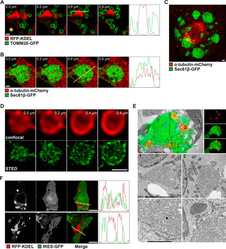
Membrane organization in SPP-induced ER clusters. A, confocal sections of HEK293T cells transfected with WT SPP, RFP-KDEL, and TOMM20-GFP as indicated. Shown are subregions of the cells next to the nuclei (*). Four frames, each from z-stacks at step sizes of 0.3 μm, are displayed. Plot profiles of signal intensities from the yellow lines superimposed on the image at 0.6 μm each are shown next to the z-stacks. The z axis represents the distance of the line, and the y axis represents the arbitrary intensities. Scale bar, 1 μm. B, confocal sections of HEK293T cells transfected with WT SPP, GFP-Sec61β, and α-tubulin-mCherry as in A. Scale bar, 1 μm. C, maximum intensity projection from confocal sections of a mitotic HEK293T cell transfected with WT SPP, GFP-Sec61β, and α-tubulin-mCherry. Scale bar, 1 μm. D, confocal (upper panel) and STED (lower panel) images of a single ER cluster of an HEK293T cell transfected with WT SPP and RFP-KDEL. Four frames, each from a z-stack at step sizes of 0.2 μm, are displayed. Scale bar, 1 μm. E, CLEM of HEK293T cells transfected with RFP-KDEL and SPP-IRES-GFP (WT). Upper left panel, low-power transmission electron micrograph of a cell of interest with fluorescence overlay (upper right panel, fluorescence light microscopy data for RFP and GFP signals). The two boxes depict ER clusters shown at higher resolution in the middle panel. The lower panel shows a closeup of the respective ER clusters. Scale bars, middle panel, 5 μm and lower panel, 1 μm. F, single confocal images from midsections of HEK293T cells transfected with WT SPP, RFP-KDEL, and GFP a few minutes after inhibitor washout (new cluster; upper panel) or 16 h after cluster formation (old cluster; lower panel). Plot profiles of signal intensities from the yellow lines superimposed on each merge image are shown. The z axis represents the distance of the line, and the y axis represents the arbitrary intensities. Scale bars, 10 μm.

