Figure 8.
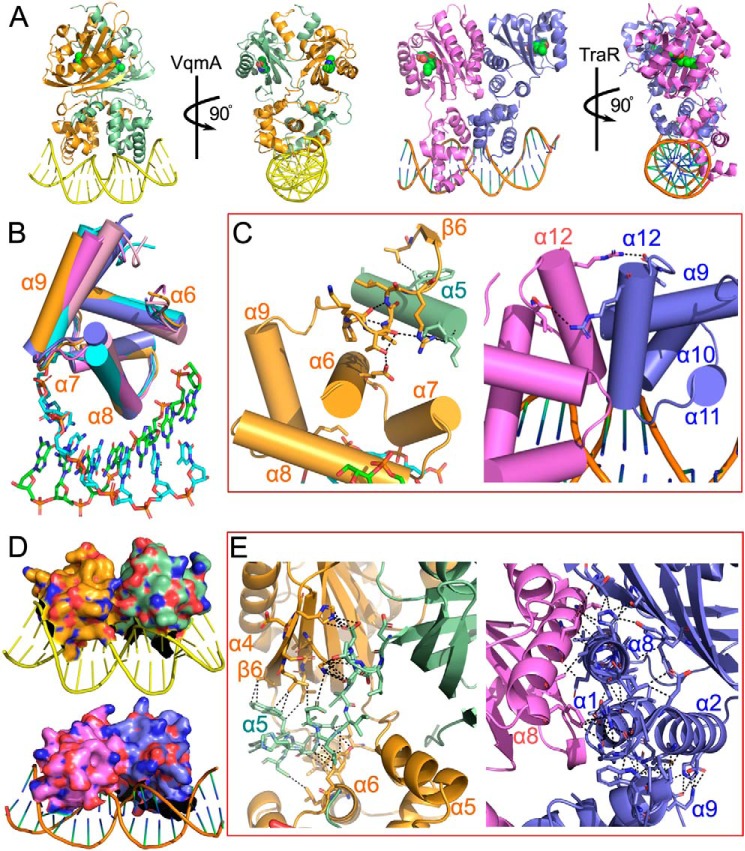
Structural analysis of VqmA and TraR. A, structure comparison of the VqmA ternary complex and TraR ternary complex. B, structure comparison of the DBD of VqmA (orange) and other DBD domains of LuxR family proteins (TraR (PDB ID 1L3L), violet; SdiA (PDB ID 4LGW), cyan; QscR (PDB ID 6CC0), pink; CviR (PDB ID 3QP5), blue). C, the necessary conditions for the stability of the last α-helix conformation in the DBD of VqmA and TraR. The interactions of the α9–β6 loop (orange) with α6 (orange) and α5 (pale green) are shown in black in VqmA. The interactions of the α12 (violet) with another α12 (blue) are shown in black in TraR. D, the comparison of dimer interfaces of DBD of VqmA and TraR. E, the conduction layers between DBD and PAS domain of VqmA and TraR. The interactions of α5 (pale green) with α6 (orange) and β-sheet (orange) are shown in black in VqmA. The interactions of α1, α2, α8 (blue) with α9 (blue) and β-sheet (blue) are shown in black in TraR.

