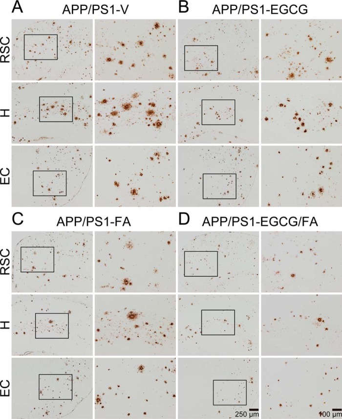
Figure 2.
Combination therapy reduces β-amyloid plaques in APP/PS1 mouse brains. A–D, representative images were taken from APP/PS1 mice that received vehicle (APP/PS1-V), EGCG (APP/PS1-EGCG), FA (APP/PS1-FA), or EGCG plus FA (APP/PS1-EGCG/FA) for 3 months starting at 12 months of age (mouse age at sacrifice = 15 months). Immunohistochemistry using an Aβ(17–24) mAb (4G8) reveals cerebral β-amyloid deposits. Brain regions shown include RSC (top), H (middle), and EC (bottom). Each right image is a higher magnification image from insets. V, vehicle.

