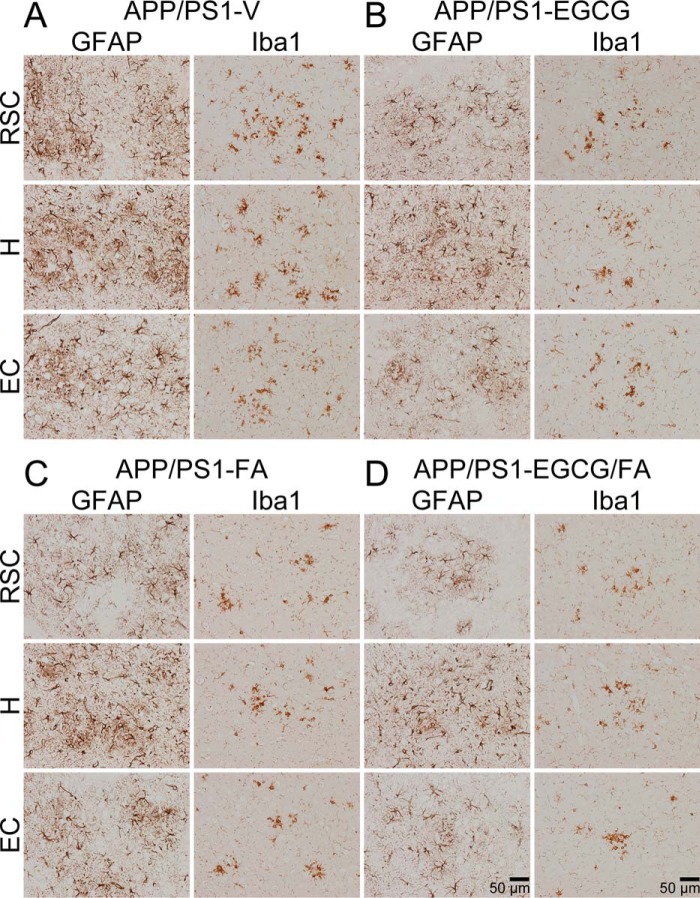
Figure 7.
Mitigated astrocytosis and microgliosis in APP/PS1-EGCG/FA mice. A–D, representative images were obtained from APP/PS1 mice that received vehicle (APP/PS1-V), EGCG (APP/PS1-EGCG), FA (APP/PS1-FA), or EGCG plus FA (APP/PS1-EGCG/FA) for 3 months starting at 12 months of age (mouse age at sacrifice = 15 months). Immunohistochemistry for GFAP and Iba1 reveals β-amyloid deposit-associated astrocytosis (left images) and microgliosis (right images). Brain regions shown include RSC (top), H (middle), and EC (bottom). V, vehicle.

