Figure 8.
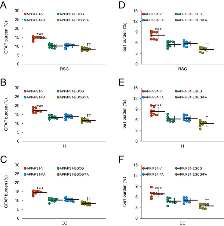
APP/PS1-EGCG/FA mice have ameliorated astrocytosis and microgliosis. Quantitative image analyses for astrocytosis (A–C) or microgliosis (D–F) burden are shown. Each brain region is indicated on the x axis. Data were obtained from APP/PS1 mice that received vehicle (APP/PS1-V, n = 8), EGCG (APP/PS1-EGCG, n = 8), FA (APP/PS1-FA, n = 8), or EGCG plus FA (APP/PS1-EGCG/FA, n = 8) for 3 months commencing at 12 months of age (mouse age at sacrifice = 15 months). Data for A–F are presented as standard deviations of the means. Statistical comparisons for A–F are within each brain region and between-groups. ***, p ≤ 0.001 for APP/PS1-V versus the other treated mice; †, p < 0.05; ††, p < 0.01 for APP/PS1-EGCG/FA versus APP/PS1-EGCG or APP/PS1-FA mice. V, vehicle.

