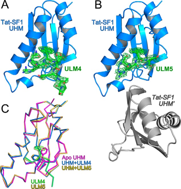
Figure 5.

Structures of Tat-SF1 UHM bound to SF3b1 ULM. A, feature-enhanced electron density maps (65) (green, 1.5 σ contour level) for SF3b1 ULM4 or B, ULM5 bound to Tat-SF1 UHM (blue). A second Tat-SF1 UHM copy that lacks bound ULM5 (Tat-SF1 UHM) is colored gray. View is rotated 180° relative to the left view of Fig. 2. C, superposed backbone traces of unliganded (magenta) Tat-SF1 UHM with ULM4-bound (blue) or ULM5-bound (yellow) UHM structures. The SF3b1 ULM4 (green) and ULM5 (yellow) are overlaid for reference. Stick diagrams show SF3b1 Trp293/Trp338 and compare Tat-SF1 Phe337 positions.
