Figure 2.

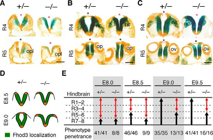
Fhod3 expression and morphological changes of the lateral neural plates of the hindbrain in Fhod3−/− embryos. A–C, lacZ staining of transverse sections of Fhod3+/− (+/−) and Fhod3−/− (−/−) embryos. Transverse sections at the level of rhombomeres 4 and 5 of lacZ-stained embryos at E8.5 (A), E9.0 (B), and E9.5 (C) are shown. Open and closed roof plates are indicated by red and black asterisks, respectively. opl, otic placode; op, otic pit; ov, otic vesicle; nc, migratory neural crest. Scale bars, 100 μm. D, schematic representation of Fhod3 expression and morphological changes of the lateral neural plates at the level of rhombomere 4. E, quantitative analysis of progression of rostral closure I. Open and closed regions are indicated by red and black arrows, respectively. The number of embryos in which the rostral zipping edge is present at each rhombomere level is shown. R1–8, rhombomeres 1–8.
