Figure 6.

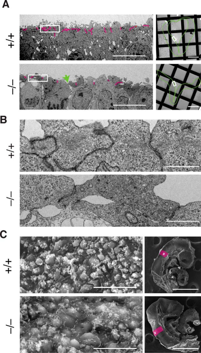
The ultrastructure of the lateral neural plate of Fhod3−/− embryos. A, transmission electron micrographs of thin sections of the apical surface of the lateral neural plates in Fhod3+/+ (+/+) and Fhod3−/− (−/−) embryos at E9.5. Cell–cell contacts (i.e. electron-dense membrane specializations) are highlighted in magenta. The green arrow indicates a loosened apical cell–cell contact. The orientation of the images in the left panels is shown as white boxes in right panels. The outlines of neural plates are indicated by green dotted lines. Scale bars, 10 μm (left panels) and 100 μm (right panels). B, examples of electron-dense membrane specializations are enlarged from images shown in A. Scale bars, 1 μm. C, scanning electron micrographs of the apical surface of the lateral neural plates in Fhod3+/+ and Fhod3−/− embryos at E9.5. The orientation of images in the left panels is shown as white boxes in the right panels. Rhombomere 4 is indicated in magenta. Scale bars, 10 μm (left panels) and 500 μm (right panels).
