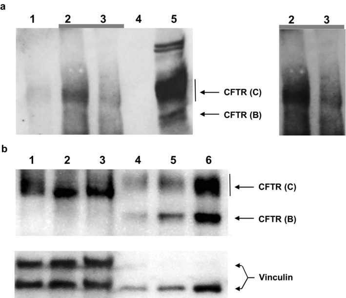
Figure 2.

CFTR immunoblot. (a) Immunodetection of CFTR (from left to right) in lysates of rectal suction biopsies by immunoprecipitation (IP) and subsequent immunoblot from a healthy non‐CF subject (lane 2) and its IP bead control (lane 1), the elder p.Phe508del/p.[Arg74;Val201Met;Asp1270Asn] compound heterozygous index case (lane 3) and its IP bead control (lane 4) and in lysates of T84 cells (lane 5). The right panel shows electronically enhanced signals of lanes 2 and 3 of the blot in order to more clearly visualize the CFTR immunoreactive signals of the biopsies. (b) Immunodetection of CFTR by immunoblot of lysates of rectal suction biopsies of two healthy non‐CF subjects (lanes 1, 3; 87 µg each) and of the younger p.Phe508del/p.[Arg74;Val201Met;Asp1270Asn] compound heterozygous index case (lane 2; 87 µg) (lane 1–3) and of non‐CF 16HBE14o cells (lane 4, 15 µg; lane 5, 35 µg; lane 6, 50 µg). Vinculin signals are provided as a western blot loading control
