Figure 4.
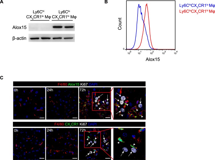
Identification of Alox15 as a specific marker for Ly6CloCX3CR1hi macrophages. (A) Western blot analysis for Alox15 expression in 24 h Ly6ChiCX3CR1lo and 72 h Ly6CloCX3CR1hi macrophages. Full-length blots are shown in Fig. S3C. (B) Flow cytometric analysis for Alox15 expression in 24 h Ly6ChiCX3CR1lo and 72 h Ly6CloCX3CR1hi macrophages. Cell populations from five mice were pooled to form one group. (C) Alox15+CX3CR1hi macrophages were in close proximity to proliferating hepatocytes. White arrows indicate proliferating hepatocytes labeled with Ki67, and arrowheads indicate macrophages labeled with F4/80 and Alox15 in WT mice (upper panel) or F4/80 in Cx3cr1GFP/+ mice (lower panel). Scale bars, 20μm. Data shown are representative of three independent experiments.

