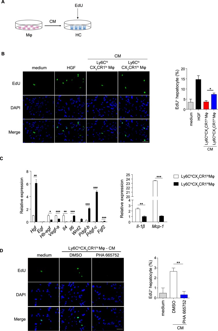
Figure 5.
Ly6CloCX3CR1hi macrophages directly accelerate hepatocyte proliferation in vitro. (A) Schematic of the experimental design. Normal hepatocytes (HCs) were co-cultured with the CM from 24 h Ly6ChiCX3CR1lo macrophages or 72 h Ly6CloCX3CR1hi macrophages. (B) Representative images of hepatocytes pulsed with EdU and the quantification of hepatocyte proliferation are shown. Scale bar, 50 μm. (C) Differential expression of the indicated genes measured by qPCR in 24 h Ly6ChiCX3CR1lo macrophages and 72 h Ly6CloCX3CR1hi macrophages, presented relative to Gapdh. (D) Hepatocytes were co-cultured with the CM from 72 h Ly6CloCX3CR1hi macrophages or supplemented with the c-Met kinase inhibitor PHA665752 (2.5 μM). Representative images of hepatocytes pulsed with EdU (left panel) and the quantification of hepatocyte proliferation (right panel) are shown. Scale bar, 50 μm. The data shown are representative of at least two independent experiments (n = 3/group). The results represent means ± SEM (*p < 0.05, **p < 0.01, ***p < 0.001).

