FIG 3.
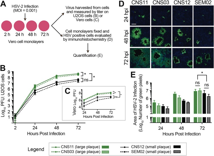
Enhanced viral cell-to-cell spread contributes to increased plaque size in culture. (A) The rates of viral spread in Vero cells were compared for representative neonatal isolates, including large-plaque formers (green) and small-plaque formers (black). Vero cell monolayers were infected at MOI = 0.001 in the presence of 0.1% human serum, which was replenished every 24 h. (B and C) Samples were harvested at each time point, and viral titers were evaluated by plaque formation on U2OS cells (B) or Vero cells (C). These data represent results from three independent experiments. Two-way ANOVA was performed followed by Tukey’s multiple-comparison test. *, P < 0.0001 at 72 h. (D) In parallel experiments, HSV-positive cells (green) were evaluated by immunofluorescence. Cell nuclei are counterstained with DAPI (blue). Scale bar = 200 μm. Images are representative of results from three independent experiments. Images of the entire 10-mm coverslips were then captured and stitched to create a composite image (see Fig. S2). (E) The total number of immunofluorescent (green) pixels was quantified for each coverslip. Two-way ANOVA was performed followed by Tukey’s multiple-comparison test. *, P < 0.05 at 72 h.

