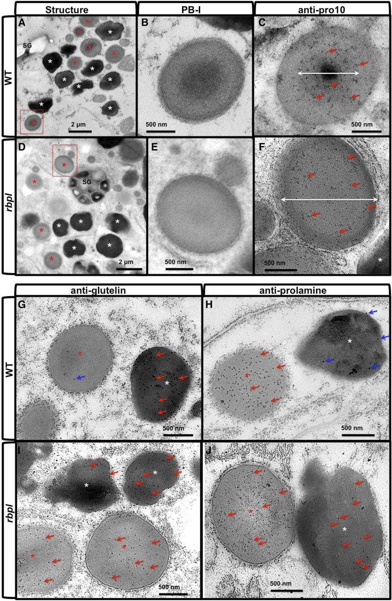
Figure 10.
PB-I structure and localization of glutelin and prolamine proteins in wild-type and rbpl line endosperm cells. A to F, Ultrastructure of PB-I in wild-type (A–C) and rbpl line (D–F) endosperm cells. B and E, Enlarged picture of the areas indicated by the red boxes in (A) and (D). C and F, Immunolabeling patterns using anti-prolamine (Pro-10) antibody and 10-nm gold particle-conjugated secondary antibody in wild type (C) and rbpl (F) PB-I, respectively. White double-headed arrows indicate the primary distribution area of Pro-10 proteins within PB-I. Scale bar = 2 μm (A and D) or 500 nm (B, C, E, and F). Red and white asterisks in (A) and (D) denote PSV and PB-I, respectively. G to J, Immunolabeling of glutelin (G, I) and prolamine (H, J) proteins using monospecific antibodies and 15-nm gold particles-conjugated secondary antibodies. G and H, wild-type. I and J, rbpl line. Red arrows denote positive gold particle labeling, and blue arrows denote slight background nonspecific labeling. Scale bar = 500 nm. WT, wild type.

