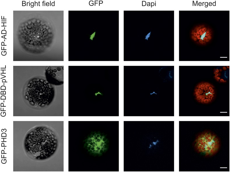
Figure 1.
Subcellular localization of the components of the oxygen-sensing machinery in Arabidopsis protoplasts. AD-HIF (Gal4 AD fusion of the HIF1α CODD domain), DBD-pVHL (Gal4 DBD fusion of the pVHL β-domain), and PHD3 DNA sequences were fused in frame with an N-terminal GFP protein sequence, expressed in Arabidopsis mesophyll protoplasts, and imaged with a confocal microscope. The signal from the GFP channel is visualized in green. 4′,6-Diamidino-2-phenylidone (Dapi) staining (1 µg mL−1) marks the nuclei (blue). In the merged image, the red color is associated with chloroplast autofluorescence. Bars = 10 µm.

