Figure 2.
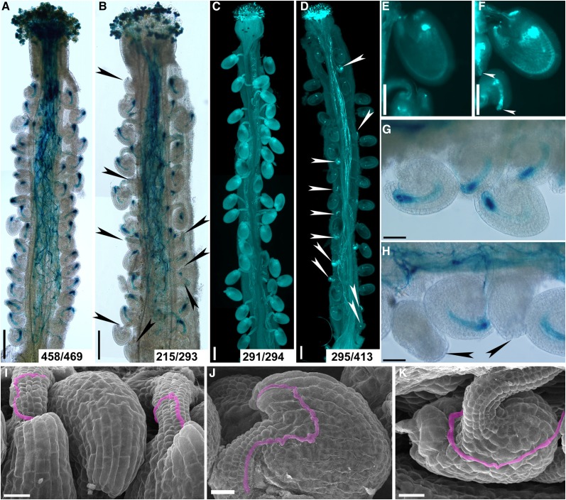
Functional loss of IMB4 compromises female-controlled pollen tube guidance. A and B, Histochemical GUS staining of wild-type (A) or imb4-1 (B) pistils at 12 HAP with ProLAT52:GUS pollen. Arrowheads point at ovules that failed to attract pollen. Two to three overlapping high-magnification images were taken for one pistil. The images were then overlaid with Photoshop (Adobe) to show the whole pistil. Numbers at the bottom: ovules targeted by a ProLAT52:GUS pollen tube versus ovules examined. C and D, Aniline blue staining of wild-type (C) or imb4-1 (D) pistils at 48 HAP with wild-type pollen. Two to three overlapping high-magnification images were taken for one pistil. The images were then overlaid with Photoshop (Adobe) to show the whole pistil. Arrowheads point at ovules that did not develop as a result of fertilization failure. Numbers at the bottom: fertilized ovules versus total ovules examined. E and F, A close-up of aniline blue stained wild-type (E) or imb4-1 (F) pistils at 48 HAP with wild-type pollen. Arrowheads point at the unfertilized ovules. G and H, A close-up of histochemical GUS stained wild-type (G) or imb4-1 (H) pistils at 12 HAP with ProLAT52:GUS pollen. Arrowheads point at the micropyles that failed to attract pollen tubes. I to K, SEMs of wild-type (I) or imb4-1 (J-K) pistils at 12 HAP with wild-type pollen. Incoming pollen tubes are highlighted in pink. A pollen tube enters through the micropyle of a normal-developing ovule of imb4-1 (J) but not of a defective ovule of imb4-1 (K). Bars = 200 µm (A to D), 100 µm (E and F), 50 µm (G and H), and 20 µm (I to K).

