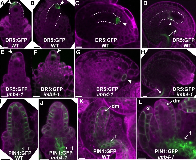
Figure 4.
Distribution of auxin maximum and PIN1 is compromised by IMB4 loss-of-function. A to H, CLSM of DR5:GFP;wild type (WT; A to D) or DR5:GFP;imb4-1 (E to H) at stage 2-III (A, E), 3-I (B, F), 3-III (C, G), or 3-V (D, H). I to L, CLSM of PIN1:GFP (I, K) or PIN1:GFP;imb4-1 (J, L) at stage 2-I (I, J) or 3-I (K, L). Arrowheads point at DR5:GFP signals. Arrows point at funiculus (f). Dotted lines highlight nucellus (A, B) or developing embryo sacs (C, D). dm, Degenerating megaspores; oi, outer integument. Cells were stained with Lysotracker red (magenta). Bars = 10 µm.

