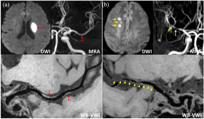
Figure 1.
Representative cases of deep-perforator infarction (DPI) (a) and non-DPI (b). (a) Diffusion-weighted imaging (DWI) with a high signal intensity lesion (red arrow) in the left basal ganglia, magnetic resonance angiography (MRA) with mild stenosis in the left middle cerebral artery (MCA) (red arrow), whole-brain vessel-wall imaging (WB-VWI) revealing focal plaques (red arrows) in the left MCA. (b) DWI showing multiple high signal intensity lesions (yellow arrow) in the right centrum ovale, multiple severe stenosis of the right MCA (yellow arrows) by MRA, WB-VWI with diffuse plaques in the right MCA (yellow arrowheads).

