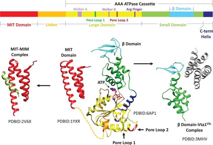
Figure 1. Domain structure of Vps4.
Top, linear sequence of eukaryotic Vps4 with domains and motifs indicated. Bottom, composite structure of Vps4 (center) with separately determined MIT [18] and ATPase cassette [22,36] structures connected by a flexible linker. The MIT domain binds the MIM sequence of ESCRT-III substrates (left) to recruit Vps4 to ESCRT-III filaments [17]. The β-domain binds the dimeric VSL domain of the Vta1 cofactor [23] (right). PDB codes of the separately determined domain and complex structures are indicated.

