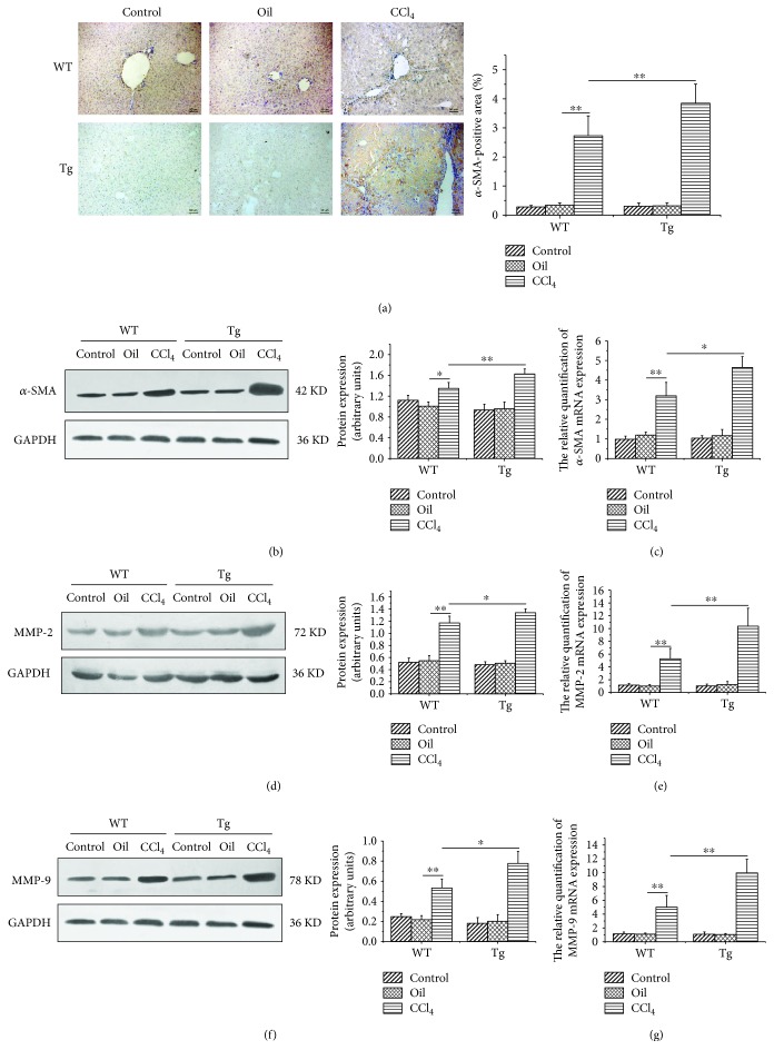
Figure 4.
Constitutive TL1A expression on myeloid cells facilitated HSC activation and the expressions of MMP-2 and MMP-9. (a–c) The α-SMA expressions in the CCl4/Tg group were markedly higher than that in the CCl4/WT group detected by immunohistochemical staining (200x), Western blot, and RT-PCR. (d, e) The MMP-2 expressions in the CCl4/Tg group were significantly increased than those in the CCl4/WT group detected by Western blot and RT-PCR. (f, g) The MMP-9 expressions in the CCl4/Tg group were notably increased than those in the CCl4/WT group detected by Western blot and RT-PCR. Data are expressed as mean ± SD, ∗p < 0.05 and ∗∗p < 0.01.

