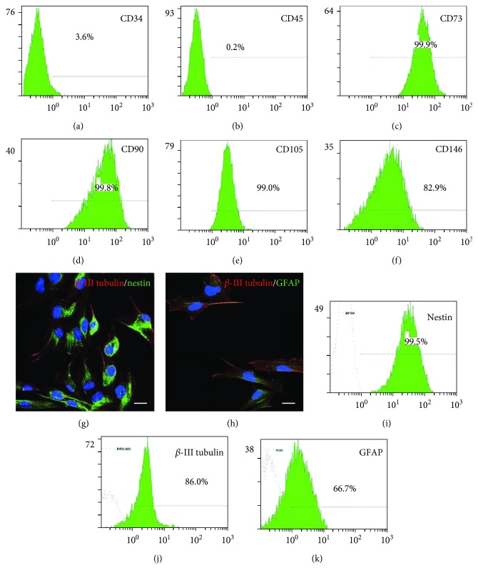
Figure 1.
Identification of SHEDs. (a)–(f) Flow cytometry plots showing that the cultured SHEDs were positive for the mesenchymal markers CD73, CD90, CD105, and CD146, but negative for the hematopoietic marker CD34 and the common leukocyte antigen CD45. (g)-(h) Representative photomicrographs of SHEDs expressing the neural stem cell markers nestin and β-III tubulin, and the glia-specific marker GFAP. Scale bars: 20 μm. (i)–(k) Flow cytometry plots showing the percentages positive for nestin, β-III tubulin, and GFAP.

