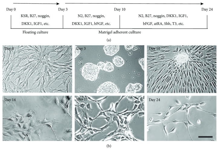
Figure 2.
Morphological changes during in vitro retinal differentiation. (a) Retinal differentiation protocol. (b) Morphological changes of SHEDs during retinal induction. On day 3, large floating neurosphere-like masses were observed. After retinal cell (factor cocktail) induction on Matrigel, the cells became larger and extended cytoplasmic processes like neurites, cross-linking with adjacent cells about 14 days post-induction. At the end of induction (day 24), cells displayed the morphology of neuron-like cells. Scale bar: 50 μm.

