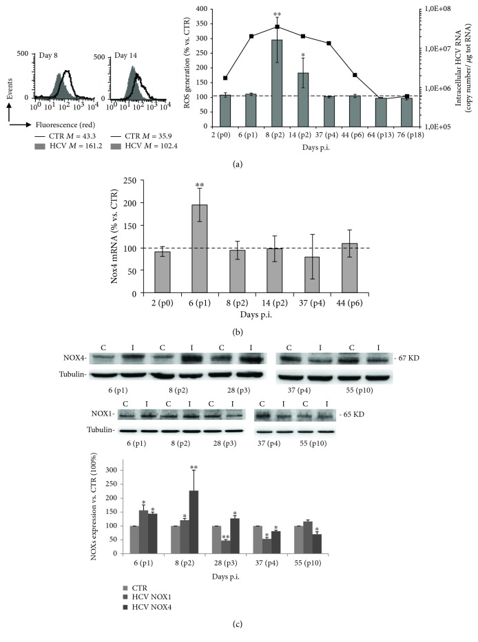
Figure 2.
HCV infection increases ROS production through the activity of NOX4. (a) Quantitative cytofluorimetric analysis of intracellular ROS production at different time points after HCV infection in cells stained with CellROX deep red. In the left panel, data obtained from the samples relative to 8 and 14 days p.i. are shown as one representative experiment out of three performed. Numbers represent the median fluorescence intensity. In the right panel, mean ± SD of the results obtained from three different experiments are reported; the results are expressed as percentage of variation vs. CTR uninfected cells considered as 100, as indicated by the dashed line. ∗P < 0.05 and ∗∗P < 0.01 vs. CTR uninfected cells. The dark line refers to the intracellular HCV genome copy number. (b) Real-time PCR assay of NOX4 isoform levels in HCV-infected Huh7.5 cells, normalized to levels in uninfected cells (CTR), indicated with the horizontal dashed line. Data shown are the means ± SD of three performed experiments ∗∗P < 0.01. (c) Western blot of uninfected (C) and HCV-infected (I) Huh7.5 cells at different times from infection, using anti-NOX4 and anti-NOX1 antibodies. Tubulin was used as a loading control. Blot is one representative experiment out of three performed. Densitometric analysis of the blots is shown. Data represent the mean ± SD of six different technical replicates; unpaired t test: ∗P < 0.05 and ∗∗P < 0.01 vs. CTR (considered as 100%).

