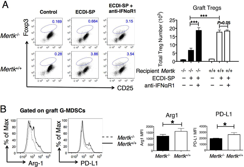
Figure 7. Cardiac allografts from Mertk−/− recipients exhibit a markedly reduced number of intra-graft CD4+CD25+Foxp3+ Tregs and an altered MDSC phenotype.

(A) Heart allografts from Mertk−/− or Mertk+/+ recipients were retrieved on post-transplant day 10 and enumerated for CD4+CD25+Foxp3+ Tregs. Grafts from control untreated recipients are shown in the left dot plots. Grafts from recipients treated with donor (BALB/c) ECDI-SP are shown in the middle dot plots. Grafts from recipients further treated with anti-IFNaR1 as described in Methods are shown in the right dot plots. Representative dot blots were all gated on CD4+ cells. The bar graph on the right shows averaged total number of intra-graft Tregs from the various groups. (B) Heart allografts from Mertk−/− or Mertk+/+ recipients treated with BALB/c ECDI-SP were retrieved on post-transplant day 10, and graft-infiltrating G-MDSCs were analyzed for expression levels of Arginase-1 and PD-L1 by FACS. Representative histograms are shown on the left. The bar graph on the right shows the averaged MFI of Arginase-1 or PD-L1 of G-MDSCs in heart allografts from Mertk−/− or Mertk+/+ recipients. Data for (A-B) were obtained and averaged from a total of eight animals per group from three independent experiments. Statistical significance was determined using t test. *P ≤ 0.05, ***P ≤ 0.001.
