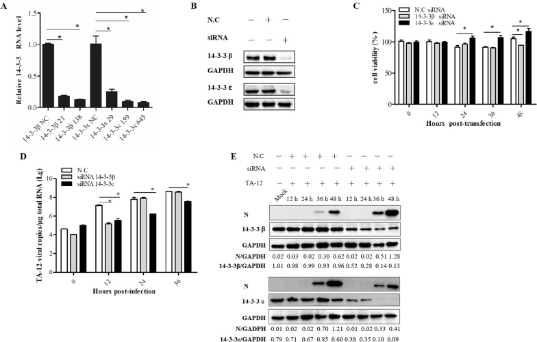
Figure 2.
Knockdown of 14-3-3ε decreases HP-PRRSV infection. A Results of qPCR for detection of 14-3-3β/ε after siRNA transfection. Marc-145 cells were transfected with siRNA. At 24 h post-transfection, the cells were collected, and total RNA was prepared for detecting the mRNA levels of 14-3-3β/ε. B Western blot analysis of 14-3-3β/ε expression after siRNA transfection. Marc-145 cells were transfected with siRNA. At 24 h post-transfection, the cells were collected, and cellular proteins were extracted for detecting 14-3-3β/ε proteins. C Marc-145 cells were transfected with siRNA and inoculated with TA-12 or mock infected with cell-culture medium at 24 h post-transfection. The mock-infected cells were collected at different time points after infection, and their viability was measured by the CCK-8 assay. D The TA-12-infected cells were collected at the same time points as the mock-infected cells and processed for total RNA extraction. Viral loads were evaluated by absolute qPCR targeting the nucleocapsid (N) gene of HP-PRRSV . E Viral proteins were evaluated by Western blot using a monoclonal antibody (6D10) targeting the PRRSV N protein. N.C: negative control. GAPDH: glyceraldehyde-3-phosphate dehydrogenase. The GAPDH gene and protein were used as internal controls for qPCR and Western blot. The density of the protein bands—measured with a fusion analysis software by using the VILBER lourmat imaging system (Fusion FX7, France)—was determined after subtracting the density of the GAPDH bands. *P value < 0.05.

