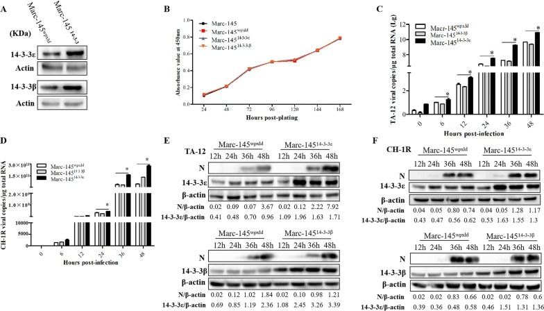
Figure 3.
Overexpression of 14-3-3ε enhances HP-PRRSV infection. A Western blot analysis of 14-3-3β/ε overexpression cells. B Cell growth kinetics. Normal Marc-145 cells and Marc-145wpxld, Marc-14514-3-3β, and Marc-14514-3-3ε cells were plated in 24-well plates. At 24, 48, 72, 96, 120, 144, and 168 h post-plating, the CCK-8 reagent was added to the cells. The cells were then incubated for 2 h at 37 °C. Cell counting was performed by measuring absorbance at 450 nm. Marc-14514-3-3β, Marc-14514-3-3ε, and Marc-145wpxld cells were infected with TA-12 and CH-1R, respectively. The infected cells were harvested at 0, 6, 12, 24, 36, and 48 hpi for RNA extraction and at 12, 24, 36, and 48 hpi for protein extraction. Viral loads were evaluated by absolute qPCR targeting the N gene of TA-12 (C) and CH-1R (D). Cellular proteins were analyzed by Western blot analysis for detecting the viral N protein of TA-12 (E) and CH-1R (F). The GAPDH protein or GAPDH gene was used as the internal control. *P value < 0.05.

