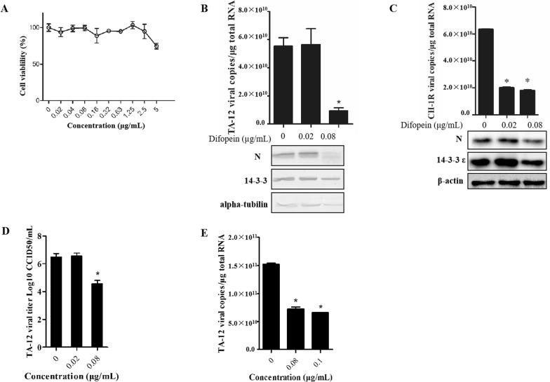
Figure 4.
Difopein decreases HP-PRRSV infection in Marc-145 cells. A The cytotoxicity of difopein was determined by the CCK-8 assay. Monolayers of Marc-145 cells in 96-well plates were treated with difopein at different concentrations for 48 h, after which the CCK-8 reagent was added to each well. After incubation for 2 h, cell viability was evaluated by measuring absorbance at 450 nm. Marc-145 cells were inoculated with TA-12 and CH-1R and incubated for 1 h, after which the medium was replaced with maintenance medium containing 0.02 or 0.08 μg/mL difopein. The cells were collected 24 h later, and total RNA and cellular proteins were extracted. Viral loads were evaluated by absolute qPCR targeting the nucleocapsid (N) gene, and viral proteins were detected by Western blot analysis of TA-12- (B) and CH-1R- (C) infected cells. D CCID50 analysis for titration of HP-PRRSV after difopein treatment. Marc-145 cells were seeded in 96-well plates. Virus supernatants were tenfold serially diluted (range 102–1010) and added to each well (at 100 μL per well) in eight repetitions. At 6 days post-infection, the numbers of cells in wells exhibiting cytopathic effects were counted, and CCID50 was calculated by the Reed–Muench method. E Marc-145 cells were infected with TA-12 and treated with 0.08 or 0.1 μg/mL difopein at 24 hpi. After incubation for another 12 h, the cells were collected for RNA extraction and qPCR analysis. *P value < 0.05.

