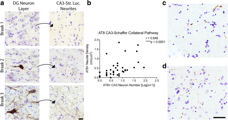
Fig. 3.
Axonal AT8 phosphorylation in the Schaffer collateral pathway occurs in the absence of CA3 cell body pathology. (a). AT8 staining in the pyramidal cell layer of CA3 and their corresponding Schaffer collateral terminal fields in the stratum radiatum of CA1 (Str. Rad.) across Braak stages. All sections were counter stained with cresyl violet. The increase in cell body staining and neuropil thread staining positively correlates with Braak staging (see Table 2). Scale bars are 25 μm. Quantification of cell body staining using total enumeration indicates the low number of cells stained in these cases. 61.5% of cases (24 of 39) displayed five or fewer cells stained in the CA3 pyramidal cell layer, with 12.8% (5 of 39) showing no observable cell body pathology (see Table 4). By comparison, 100% of cases displayed axonal AT8 staining in the CA1 Str. Rad. (b). Spearman correlation analysis of AT8+ axonal density and cell body number in the Schaffer collateral pathway. A strong, positive correlation (r = 0.648, p ≤ 0.0001) indicates an increase in axonal pathology as cell body pathology increases. (c-d). Representative images from cases with sparse (c) and dense (d) AT8+ Schaffer collateral axons in cases lacking cell body pathology demonstrate the extent of axonal pathology that can occur prior to observable somatodendritic pathology. Scale bars are 50 μm

