Fig. 7.
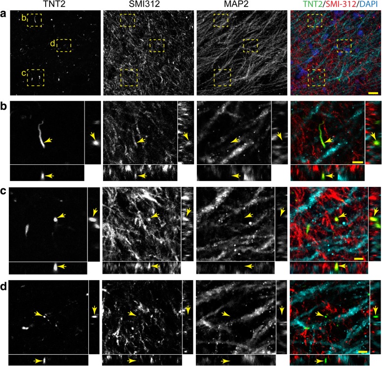
TNT2+ tau neurite pathology colocalizes with the axonal marker SMI-312 in the Schaffer collateral pathway. (a-d). Representative images of TNT2 (green), SMI-312 (red) and MAP2 (cyan) triple labeling immunofluorescence staining in the Schaffer collaterals of the hippocampus (merged image in a includes DAPI nuclear counter stain). (a) A low magnification image shows axonal (SMI-312), dendritic (MAP2) and tau+ neurites (TNT2) in the CA1 Str. Rad. region of the hippocampus in a Braak stage III case. (b and c) Cross-sectional analysis of z-stack images (60x magnification 0.5 μm step size) demonstrate that TNT2+ neurites colocalize with SMI-312 (arrows). (d) Cross-sectional analysis of z-stack images show that some TNT2+ neurites do not colocalize with SMI-312 or MAP2. Notably, little to no colocalization was observed between TNT2+ neurites and MAP2. Scale bars are 20 μm for a and 5 μm for b-d. AT8+ tau neurite pathology showed similar colocalization with SMI-312, not MAP2 in the Schaffer collateral pathway (see Additional file 6: Figure S6)

