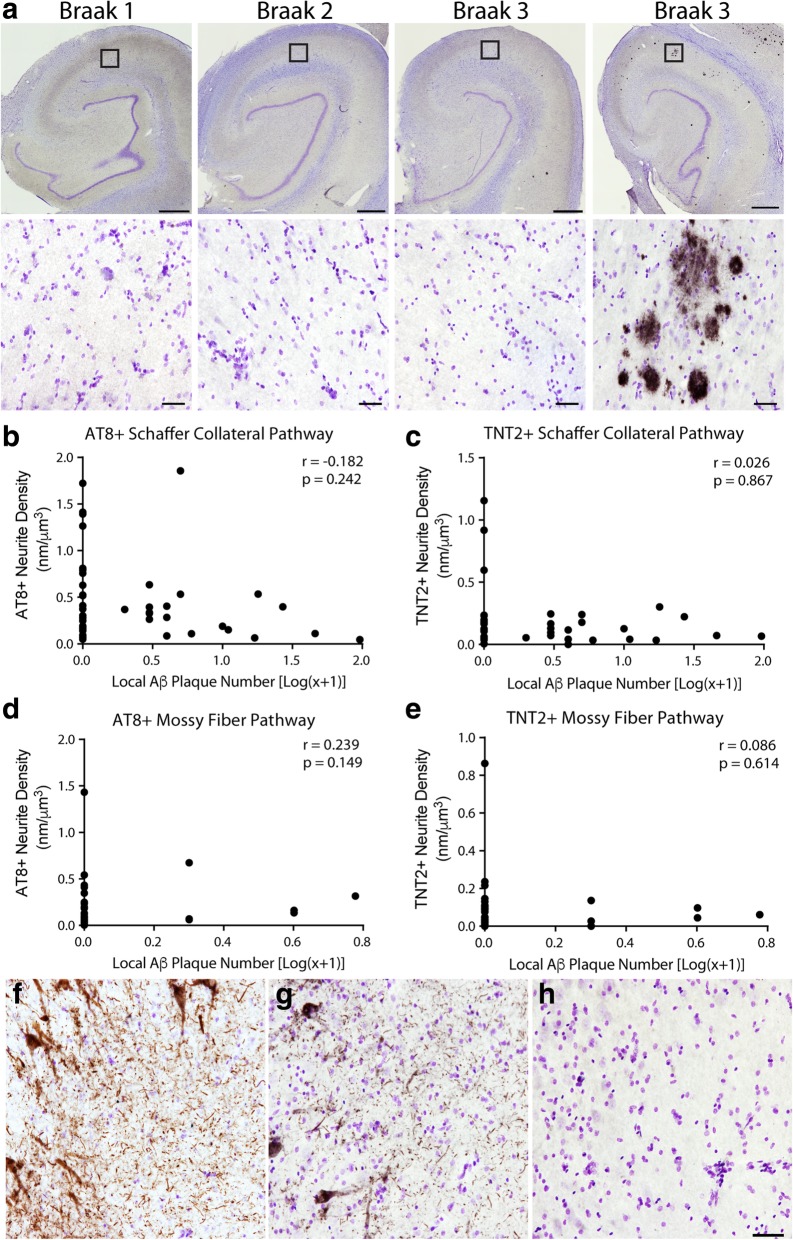
Fig. 8.
Local tau pathology is observed in the absence of local amyloid-β pathologies. (a). Representative hippocampi across Braak stages stained with MOAB2. Note the rarity of MOAB2+ plaques in the hippocampus and in the Schaffer collateral terminal region of the CA1 Str. Rad. (similar lack of Aβ pathology occurred in the mossy fiber terminal region of the Str. Luc.). A representative image of the relatively rare cases containing a significant amyloid load in the hippocampus is shown to confirm the effective labeling of Aβ plaques with MOAB2 antibody. Boxes indicate the location of higher magnification images. In the mossy fiber pathway, 84.2% of cases contained zero plaques in the CA3 Str. Luc. and 59% of cases contained zero plaques in the DG (see Table 7). In, the Schaffer collateral pathway, 55.8% of cases contained zero plaques in the CA1 Str. Rad. and 78.9% of cases contained zero plaques in the CA3 pyramidal cell layer (see Table 7). Scale bars are 1 mm in upper images and 50 μm in lower images. (b, c). No significant correlation was observed in the Schaffer collateral pathway between the presence of local Aβ plaques and local AT8+ staining (b, Spearman r = − 0.182, p = 0.242) or TNT2+ staining (c, Spearman r = 0.026, p = 0.867). (d, e). No significant correlation was observed in the mossy fiber pathway between the presence of local Aβ plaques and local AT8+ staining (d, Spearman r = 0.239, p = 0.149) or TNT2+ staining (e, Spearman r = 0.086, p = 0.614). (f, h). Representative images of AT8 (f), TNT2 (g), and MOAB2 (h) staining in the same location within the CA3 region from the same case illustrates the robust tau pathology possible in the absence of detectable Aβ pathology. It is noteworthy that no cases contained intraneuronal MOAB-2 staining for soluble monomeric or oligomeric Aβ species. The only Aβ pathologies observed in these hippocampal regions were extracellular plaques. Scale bar is 50 μm and applies to panels f, g, and h

