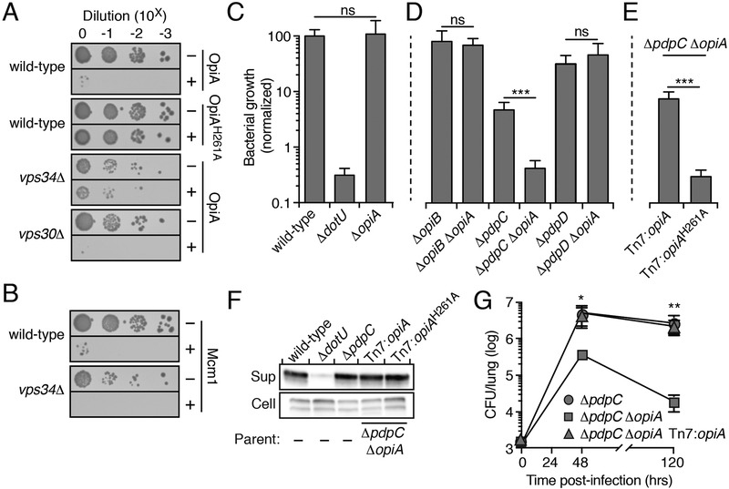
Figure 3.
The PI3K activity of OpiA leads to toxicity when expressed in yeast and contributes to F. novicida growth in vivo
(A and B) Growth resulting from serial dilution of the indicated yeast strains carrying plasmids to express the noted OpiA alleles (A) or Mcm1(B) plated on inducing (+) or non-inducing (−) media.
(C-E) Growth at 24hrs of the indicated strains of F. novicida in RAW 264.7 cells, normalized to level of growth by the wild-type strain (100%). Data are shown as the mean ± s.d. Asterisks represent statistically significant differences (Student’s t-test; ***p ≤ 0.0001).
(F) Western blot analysis of OpiA or OpiAH261A in the supernatant (sup) and cellular (cell) fractions of the indicated strains of F. novicida grown in the presence of 5% (w/v) potassium chloride.
(G) Quantification of the bacterial burden in the lungs of mice infected via aerosolization with the indicated strains of F. novicida. Data are shown as the mean ± s.d. and asterisks represent statically significant differences (ANOVA; *p ≤ 0.05, **p ≤ 0.001).

