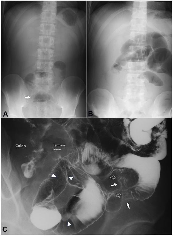
Figure 2. Plain abdominal radiographs on admission (A) and 13 days after the first infusion of infliximab (B). Solitary air-fluid level is indicated by an arrow (A). Multiple air-fluid levels are seen (B); C – Picture of the enteroclysis. Two stenotic portions are observed in the distal part of the ileum (white arrows). At the sites of stenosis, white soft shadows are seen (hollowed arrows) indicating active ulcers. Pre-stenotic dilatation is not observed. Longitudinal ulcers are observed (arrowheads).

